It’s over 130 years since Wilhelm Conrad Röntgen discovered x-rays. They were used in medical diagnosis almost immediately, but veterinary radiology was slower to take off. Here in Australia, x-rays weren’t part of standard veterinary diagnosis till the 1970s and ‘80s.
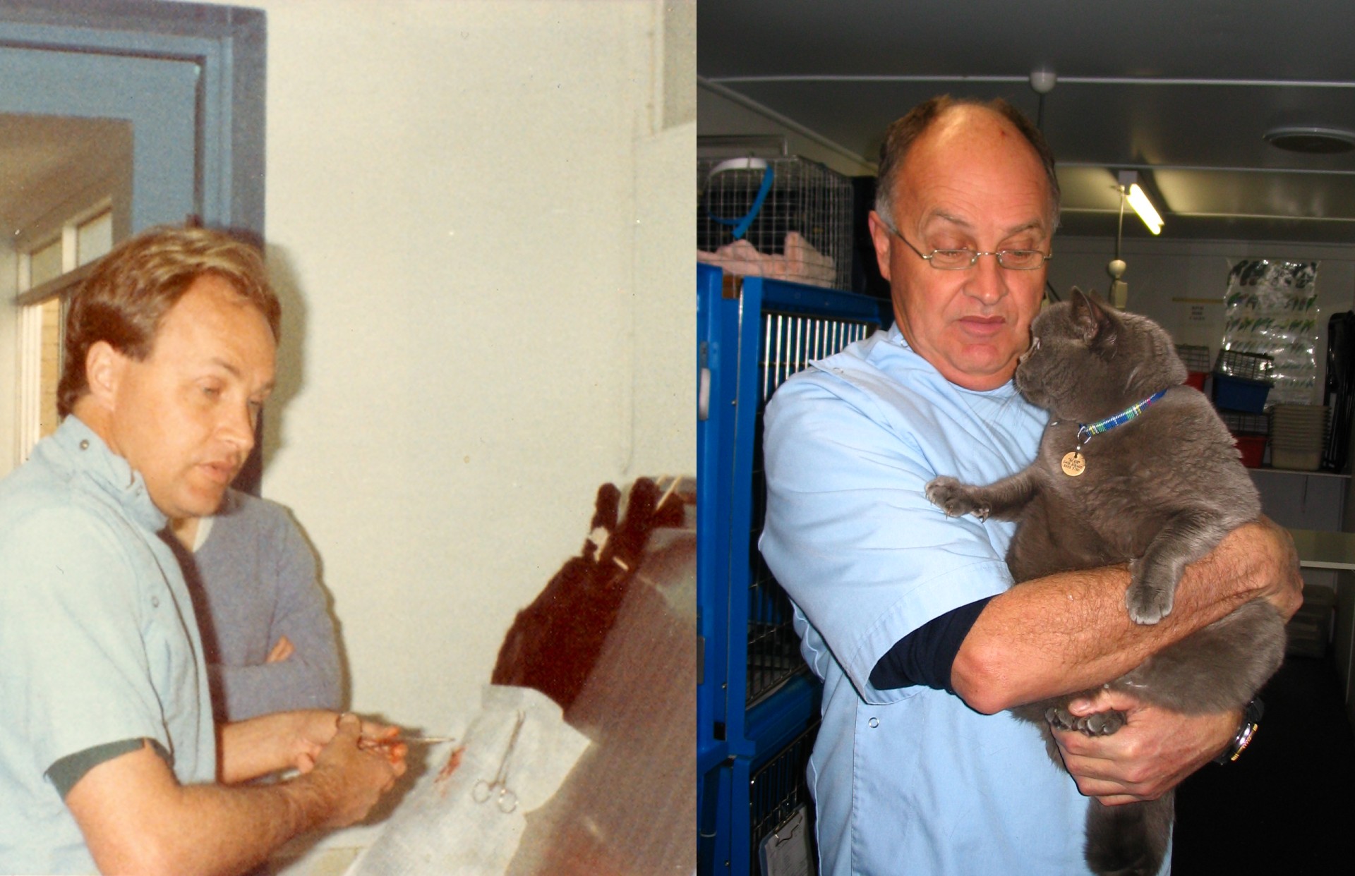
We spoke to Tony Mosman, former partner of Bondi Junction Vets, about his early years in practice. It’s amazing how much has changed, in technology generally and in veterinary radiology in particular.
When did you start practising as a vet?
I graduated back in 1971, and joined a practice in Cowra, then went out to West Wyalong, 300 miles due west of Sydney. (500km in modern measures.)
How was it different from today?
The first thing is that I was on call 24×7, seven days a week.
This was long before the days of the mobile phone. In fact, out in West Wyalong, we had the last manual telephone exchange in NSW. The operators had to physically plug the wires in to connect us.
Somehow, they always knew where I was. I might be at the pub or the club or on someone’s property, but they always found me. I used to tell them where I was going, too. One thing about a manual exchange was that you always got to speak to the operator.
The vet practice was on number 707, and the doctor was on number 717. Sometimes an operator would mix the wires up. I might get a call about a person with a broken arm, or the doctor would be asked to treat a horse. We always sorted things out, but it was amusing. article.)
How long were you there?
I was only out west for a year or so, then I went to England for 4 years. I spent time as a locum all over – everywhere from Hull to Truro to Liverpool – and travelled in summer and spent winters in Austria as a ski instructor. But in 1975 I came back for my sister’s wedding, and that’s when I started at Bondi Junction Vet.
Tell us about the early days of the Bondi Junction practice.
The practice was built by Norman Larkin in 1934 and was the first purpose-built veterinary practice in Sydney. When I joined, the practice was owned by Bob Steel. The downstairs area was a vet hospital, and upstairs was a 3 bedroom flat. Veterinary students lived in the flat and were employed to care for hospital patients and answer after hours phone calls and door knocks, then to get in touch with the duty veterinarian of that night for advice or further treatment as required. I lived in Surry Hills, then in Randwick. There was a lot less traffic then, and I could get from home to the practice in 10 minutes. Completely different from those sometimes 150 km each way house calls I used to do at West Wyalong.
Tell us about veterinary radiology at the practice when you started.
At the start, we didn’t have a specialist x-ray area. There was just an open space in the hospital cage area.
The equipment was very different too. X-rays were directed from the head of the unit down to the x-ray table via a 30cm metal cone. To ensure the x-ray beam was directed in the right and correctly focused on the patient. We used a lead weight on a string from the cone. If the weight was directly above the animal, then the alignment was correct.
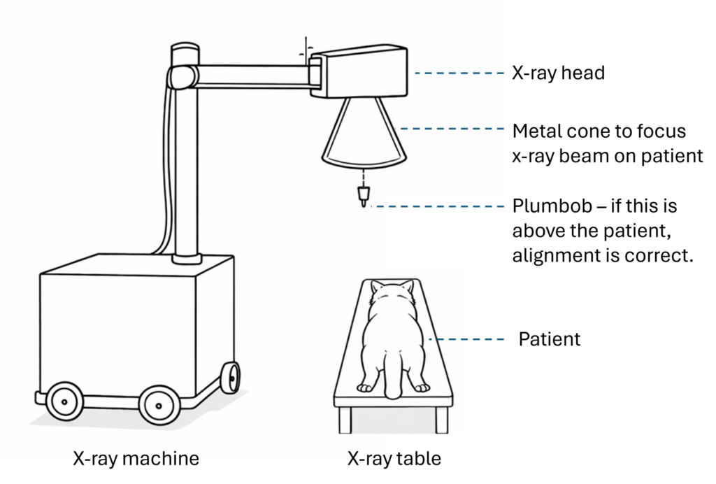
It wasn’t so bad when the animals were anaesthetised, but sometimes they weren’t and we’d have a lively time with one vet or vet nurse holding the patient while the other one operated the x-ray equipment.

Developing x-rays was a challenge too.
There was no digital radiography. Everything was film, which we had to take to the dark room to developOur dark room was in the basement, under the hospital. We had to go outside behind the hospital to access it. No fun when it was raining and we were carrying x-ray plates. The films were taken out of the x-ray cassettes and clipped onto metal frames. Then it was 3 minutes in the developer fluid, water to rinse off and 5 minutes in the fixer, then back outside and into the hospital to look at the results on our wall viewer.
Sometimes the x-ray quality was sufficient but if not we’d have to do the whole thing all over again!
When did you start working with equipment from Radincon?
It’s feels as if I’ve known Jon for ever. We used to get x-ray gear from a generalist vet supplier company, but Jon and the Radincon team were more knowledgeable about radiology and imaging in general. They were always very helpful, which made everything easy.
We’ve upgraded our x-ray equipment with Radincon 4 times over the years. We started some time back in the late 70s, so that shows how well good equipment can last!
What changes have you seen over time?
The first change we all loved wasn’t the technology. It was when we moved the dark room upstairs under cover!
The next step was installing an automatic film developer, which saved a lot of frustration.
From film to digital
A major technological change was moving from film to digital radiography. We were an early adopter back in the late 1980s. All that rushing off to dark rooms and waiting to see if the x-rays were any good just disappeared – fantastic!
It took away the need to handle and store film too. You don’t realise how many x-rays a practice takes over the years, and how much space the film takes up. We had to keep every film for at least two years, and some of it we kept much longer. In fact, when we cleared out the old dark room under the house 6 months ago, there were several wheelbarrow loads of film still in there.
The cloud – storing and sharing images
Our digital images were all stored locally at first. We had a computer running MS-DOS, and whoever needed to see the images had to use that computer. It wasn’t like nowadays, when you can share images with a specialist anywhere in the world in no time.
Image quality
Over the years, I’ve seen veterinary radiology images get better and better. Once upon a time, we could see the bones, but the fine detail wasn’t there, and soft tissue was hard to distinguish too.
We’ve always seen a fair number of animals because they swallowed something. Metal objects were easy to spot, but plastic items, like the soft tissues, didn’t show up very well. Sometimes all we could see was distortion of the bowel, or a black space where gas had built up behind the obstruction. We had to guess what was going on based on that.
Nowadays, you can see so much more, including those plastics, and you can seem them more clearly.
Safety considerations
Another area I think has improved is compliance with x-ray safety. We always knew about health risks, and did our best with lead gloves and aprons, but not were not always aware of x-ray radiation exposure, though we did wear radiation exposure badges. But we didn’t always take them seriously. In one practice I once worked in, the general assistant would be holding animals while we took x-rays, but not wearing any apron, collar or gloves at all.
Overall, veterinary radiology has come a long way. The images are better, even with lower radiation doses. And images are available easily, not just for the vet in the practice, but to share with specialists. It all contributes to better diagnosis, meaning more options for treatment.
Any final thoughts or advice for other vets?
I’d just say, try to enjoy it. There’s a lot of variety, and I always loved animals, so it was no problem for me to spend 55 years in practice. I didn’t get bored as you never knew what or who was coming next.
You do have to work on relationships with the animals you treat, and with their owners. I’ve dealt with everyone – from lords and ladies, film, TV and sporting stars, even a Prime Minister’s wife, right down to those sleeping rough. Most of them are wonderful and I enjoyed our friendly relationships. About 5% make things difficult. Try not to worry about them.
Focus on the other 95% of your clients who are good people. Connect with them and with the animals. It’s what makes the job worthwhile.